Chordoma
What is a Chordoma?
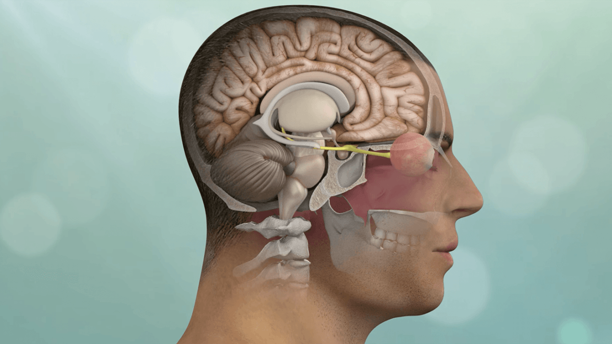
A chordoma is a rare type of malignant tumor that arises from remnants of the notochord, a structure that is present during early fetal development and provides support for the developing spine. Chordomas typically occur in the bones of the skull base, spine, or sacrum (the triangular bone at the base of the spine). These tumors grow slowly but are locally invasive and have a tendency to recur after treatment.
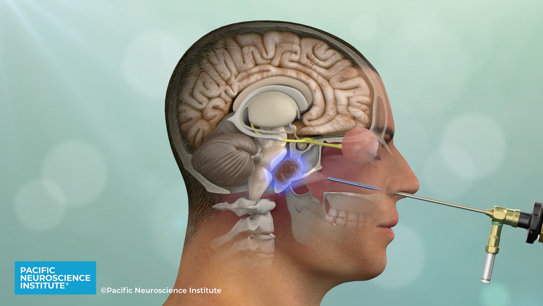
Clival chordomas can be considered as brain tumors or pituitary tumors. Clival chordomas are locally invasive tumors of the midline skull base. Chordomas are locally invasive slow-growing malignant tumors that arise from the remnant of the primitive notochord. They occur most commonly in the skull base (clivus) and lower spine. Approximately 40% of chordomas and chondrosarcomas arise in the clivus (directly below the sella turcica and pituitary gland).
Treatment for Chordomas
Endoscopic endonasal surgery is the optimal initial treatment for most Clival Chordomas
Clival chordomas are ideally treated with maximal safe surgical removal followed by focused radiotherapy. Fortunately, the majority of clival chordomas can be removed via the nose using an endoscopic endonasal approach. Occasionally, these tumors require multiple keyhole operations including the retrosigmoid and eyebrow craniotomy approaches.
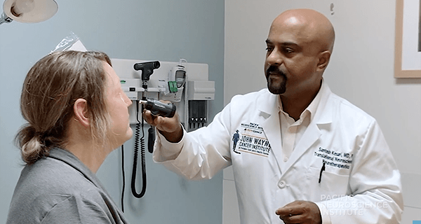
At PNI, we have one of the world’s largest experiences with endonasal surgery for clival chordomas including patients who have had prior surgery and radiation. By incorporating cutting edge technology and instrumentation with proven surgical experience, our highly collaborative team provides safe, less invasive and effective chordoma surgery.
Given that many chordomas can be resistant to both surgery and radiosurgery, tumor genomic profiling is routinely performed in our chordoma patients to find alternative targeted therapies. We also have a protocol with pemetrexed (Alimpta) for patients with chordoma which we can offer for some patients.
Chordoma Overview
Chordoma are locally invasive slow-growing malignant tumors that arise from the remnant of the primitive notochord. They occur most commonly in the skull base (clivus) and lower spine. Approximately 40% of chordomas and chondrosarcomas arise in the clivus (directly below the sella turcica and pituitary gland).
Chordomas can behave quite aggressively and are often extensively invasive into surrounding skull base structures. A small subset of patients (~10%) may develop metastases to distant sites beyond their site of origin.
Symptoms of Chordoma
The most common presenting symptom of a clival chordoma is double vision.
Less common symptoms may include:
- Facial numbness or tingling
- Loss of vision
- Hearing loss
- Difficulty swallowing
- Coordination
- Motor weakness
- Nasal congestion
- Pituitary gland dysfunction
- Headache
Diagnosis of Chordoma
These skull base tumors are best diagnosed by MRI and CT scans which will clearly show the extent of tumor and bony destruction.
Focused MRIs of the pituitary region, sinuses, temporal bones or internal auditory canals may be indicated to obtain better anatomical detail of a chordoma.
Other tests may also be needed prior to surgery such as angiography (typically now performed as a CT angiogram or an MR angiogram), visual field tests, an audiogram or pituitary hormonal tests.
Treatment for Chordoma
The initial treatment for a clival chordoma is maximal safe surgical removal.
Given their midline location, the great majority of clival chordomas and chondrosarcomas are best removed via an endoscopic endonasal approach.
However, some extensive and/or laterally placed chordomas may require different skull base approaches. Because chordomas typically invade the bone and dura of the skull base as well as cavernous sinus area, complete surgical resection is often not possible and continued growth of a residual tumor is common.
Extensive surgery can certainly improve long term survival but over-aggressive tumor removal can be associated with significant neurological complications.
After maximal safe removal, most chordoma patients will require Stereotactic Radiotherapy (SRT), stereotactic radiosurgery (SRS) or proton beam radiation. Chemotherapy is generally considered ineffective for treating chordomas although clinical trials of some experimental chemotherapies and immunotherapies are available. At Pacific Neuroscience Institute our innovative precision neuro-oncology approach has been successful in controlling tumor growth in many cases where surgical options have been exhausted.
Multidisciplinary Chordoma Specialist Team
Videos Related to Chordoma
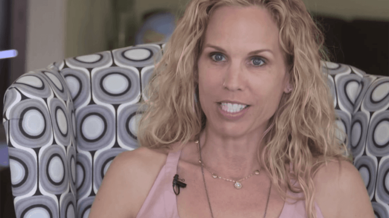
 Andrea’s Story – Clival Chordoma
Experience an actual account of Clival Chordoma.
Andrea’s Story – Clival Chordoma
Experience an actual account of Clival Chordoma.
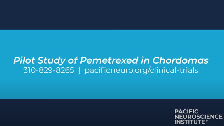
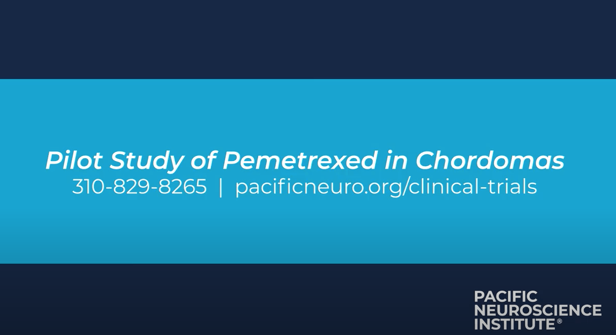 Pilot Study of Pemetrexed in Chordomas
Pacific Neuroscience Institute is now recruiting patients with chordoma for a new clinical trial at the Pacific Brain Tumor Center. Watch the video and learn about the trial.
Pilot Study of Pemetrexed in Chordomas
Pacific Neuroscience Institute is now recruiting patients with chordoma for a new clinical trial at the Pacific Brain Tumor Center. Watch the video and learn about the trial.
 Clival Chordoma Clinical Trial: Kimberly’s Story
Lean about how a patient with a recurring clival chordoma brain tumor found Dr. Santosh Kesari at Pacific Neuroscience Institute, and the clinical trial protocol where she received treatment with…
Clival Chordoma Clinical Trial: Kimberly’s Story
Lean about how a patient with a recurring clival chordoma brain tumor found Dr. Santosh Kesari at Pacific Neuroscience Institute, and the clinical trial protocol where she received treatment with…
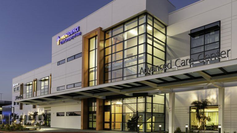
 The Pacific Brain Tumor Center
 Nuestro Brain Tumor Center
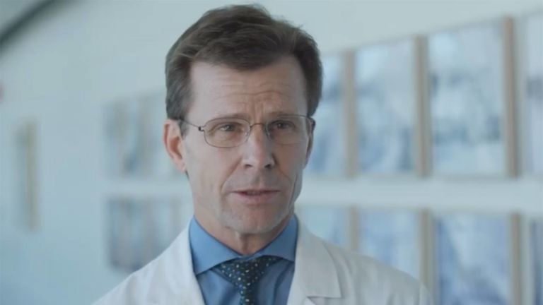
 Meet Dr. Daniel F. Kelly
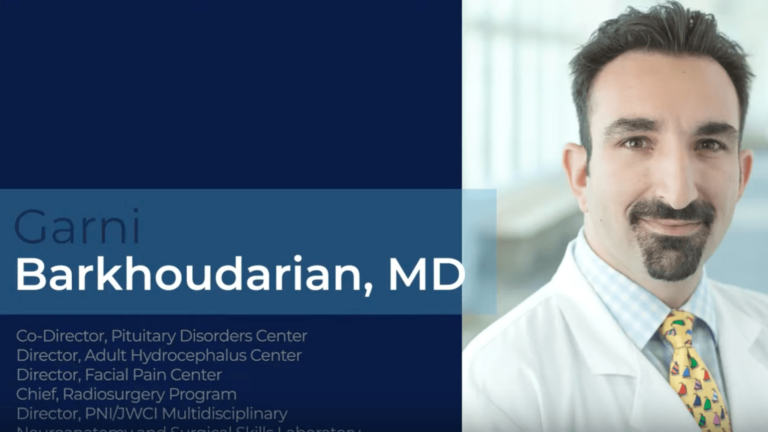
 Meet Dr. Garni Barkhoudarian
Dr. Barkhoudarian is the Co-Director Pituitary Disorders Center, Neurosurgery; Director, Adult Hydrocephalus Center; Director, Facial Pain Center; Chief, Radiosurgery Program; Director, JWCI / PNI Microdissection Anatomy Laboratory. He is a…
Meet Dr. Garni Barkhoudarian
Dr. Barkhoudarian is the Co-Director Pituitary Disorders Center, Neurosurgery; Director, Adult Hydrocephalus Center; Director, Facial Pain Center; Chief, Radiosurgery Program; Director, JWCI / PNI Microdissection Anatomy Laboratory. He is a…
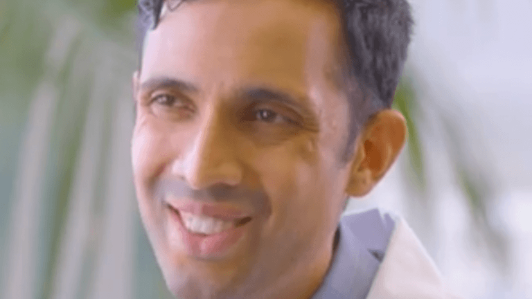
 Meet Dr. Adi Iyer
 Meet Dr. Nouzhan Sehati
Dr. Sehati, is a board-certified neurosurgeon who specializes the treatment of a wide variety of brain, spine and spinal cord disorders at Pacific Neuroscience Institute. His expertise is in surgical…
Meet Dr. Nouzhan Sehati
Dr. Sehati, is a board-certified neurosurgeon who specializes the treatment of a wide variety of brain, spine and spinal cord disorders at Pacific Neuroscience Institute. His expertise is in surgical…
 Meet Dr. Walavan Sivakumar
Dr. Walavan Sivakumar is a fellowship-trained neurosurgeon with a focus on skull base and minimally invasive and endoscopic neursosurgery. Selected as a multiple year recipient of the SuperDoctor Rising Stars…
Meet Dr. Walavan Sivakumar
Dr. Walavan Sivakumar is a fellowship-trained neurosurgeon with a focus on skull base and minimally invasive and endoscopic neursosurgery. Selected as a multiple year recipient of the SuperDoctor Rising Stars…
 Meet Dr. Howard R. Krauss
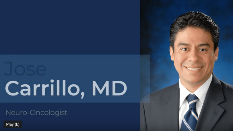
 Meet Dr. Carrillo
Dr. Carrillo is a board-certified neurologist and neuro-oncologist specializing in the diagnosis and treatment of primary and secondary brain tumors, including glioblastoma multiforme, primary CNS lymphoma, meningioma, and brain metastases,…
Meet Dr. Carrillo
Dr. Carrillo is a board-certified neurologist and neuro-oncologist specializing in the diagnosis and treatment of primary and secondary brain tumors, including glioblastoma multiforme, primary CNS lymphoma, meningioma, and brain metastases,…
 Meet Dr. Santosh Kesari
 Meet Dr. Sharma
Meet Dr. Akanksha Sharma, who is board certified in neurology, neuro-oncology and palliative medicine. She is experienced in and provides unique perspective on the treatment of primary/metastatic brain tumors, neurological…
Meet Dr. Sharma
Meet Dr. Akanksha Sharma, who is board certified in neurology, neuro-oncology and palliative medicine. She is experienced in and provides unique perspective on the treatment of primary/metastatic brain tumors, neurological…
 Meet Dr. Wagle
Naveed Wagle, MD, has specialized training and research experience in the treatment of cancer of the central and peripheral nervous system. Dr. Wagle treats patients with primary and metastatic brain…
Meet Dr. Wagle
Naveed Wagle, MD, has specialized training and research experience in the treatment of cancer of the central and peripheral nervous system. Dr. Wagle treats patients with primary and metastatic brain…
 Keyhole Brain Tumor and Skull Base Surgery
Andrea’s Story
Clinical Trials Connections: Pemetrexed for the treatment of Chordoma
Clival Chordoma Clinical Trial: Kimberly’s Story

The Pacific Brain Tumor Center
Nuestro Brain Tumor Center
Meet Dr. Daniel F. Kelly
Meet Dr. Barkhoudarian
Meet Dr. Adi Iyer
Meet Dr. Nouzhan Sehati

Meet Dr. Walavan Sivakumar
Meet Dr. Howard R. Krauss
Meet Dr. Carrillo

Meet Dr. Santosh Kesari
Meet Dr. Sharma

Meet Dr. Wagle

Keyhole Brain Tumor and Skull Base Surgery
Stories Related to Chordoma

Brain Tumor Clinic Locations
The Brain Tumor Center’s state-of-the-art facilities are located at:
Pacific Neuroscience Institute
2125 Arizona Ave., Santa Monica, CA 90404
310-582-7450
PNI – South Bay
5215 Torrance Blvd., #300, Torrance, CA 90503
424-212-5361
Providence Saint John’s Health Center
2200 Santa Monica Blvd., Santa Monica, CA 90404
310-829-8265
PNI – Burbank
501 S. Buena Vista St., Burbank, CA 90505
818-847-6049
Useful Links
- Brain tumor clinical trials
- Patient guide to endoscopic endonasal surgery
- Brain tumor fact sheet
- Pituitary disorders fact sheet
- Chordoma Foundation